 Image 1 of 19

 Image 2 of 19

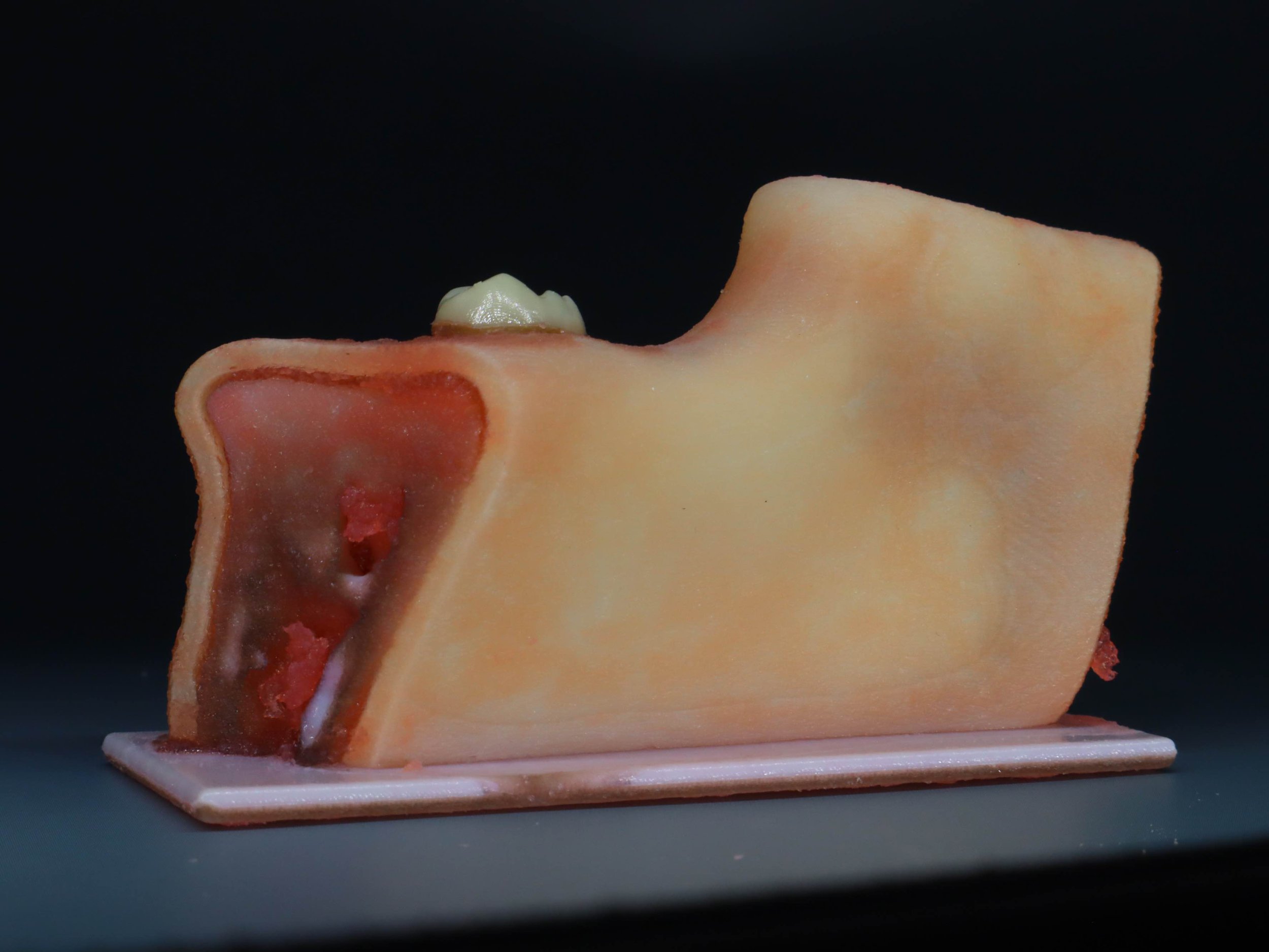 Image 3 of 19

 Image 4 of 19

 Image 5 of 19

 Image 6 of 19

 Image 7 of 19

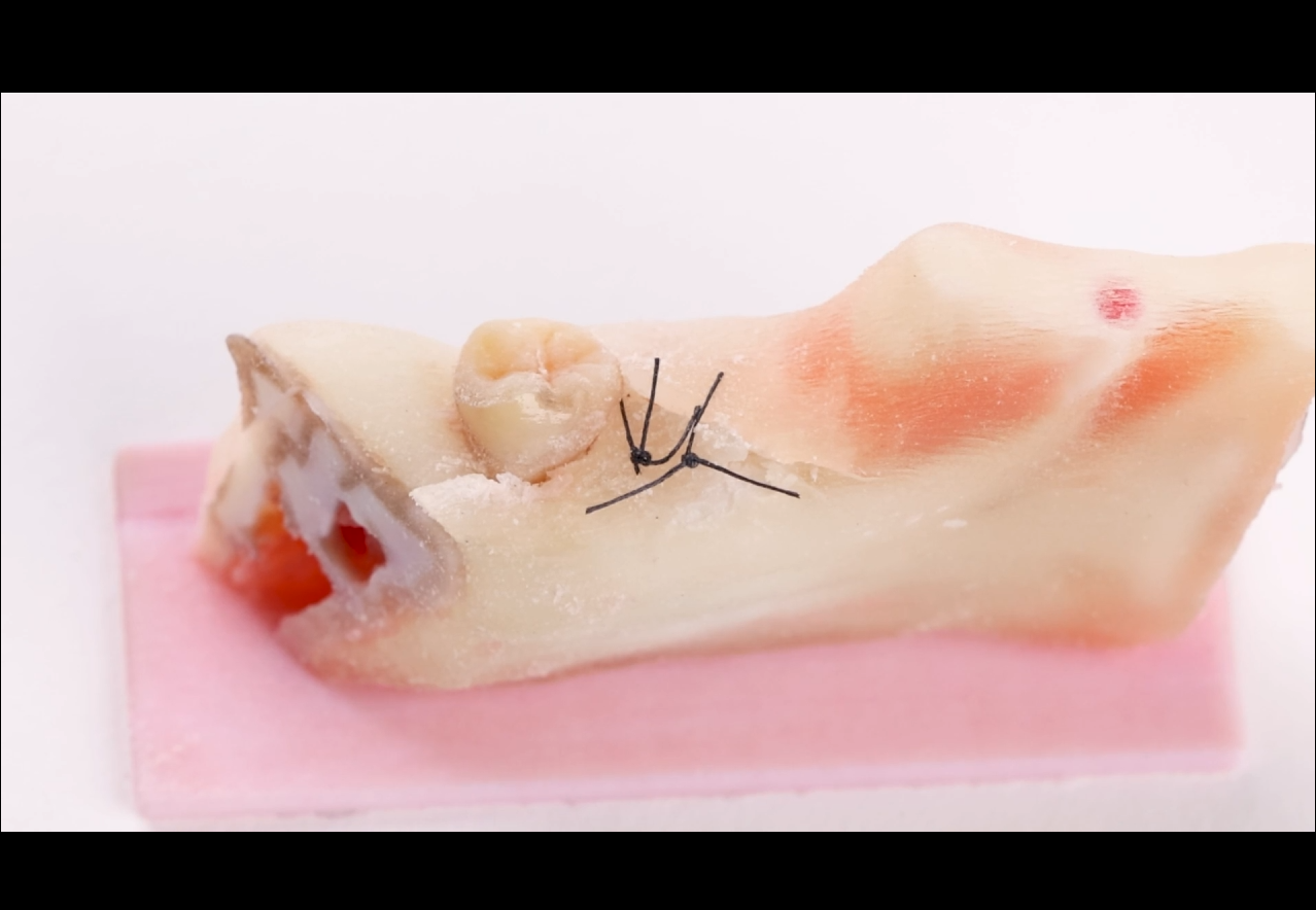 Image 8 of 19

 Image 9 of 19

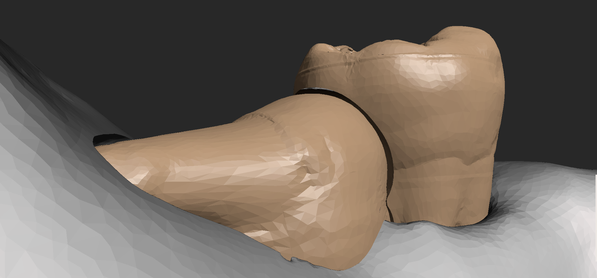 Image 10 of 19

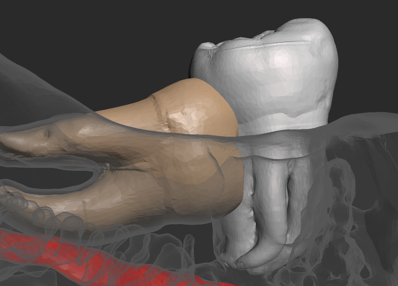 Image 11 of 19

 Image 12 of 19

 Image 13 of 19

 Image 14 of 19

 Image 15 of 19

 Image 16 of 19

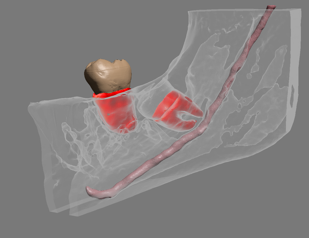 Image 17 of 19

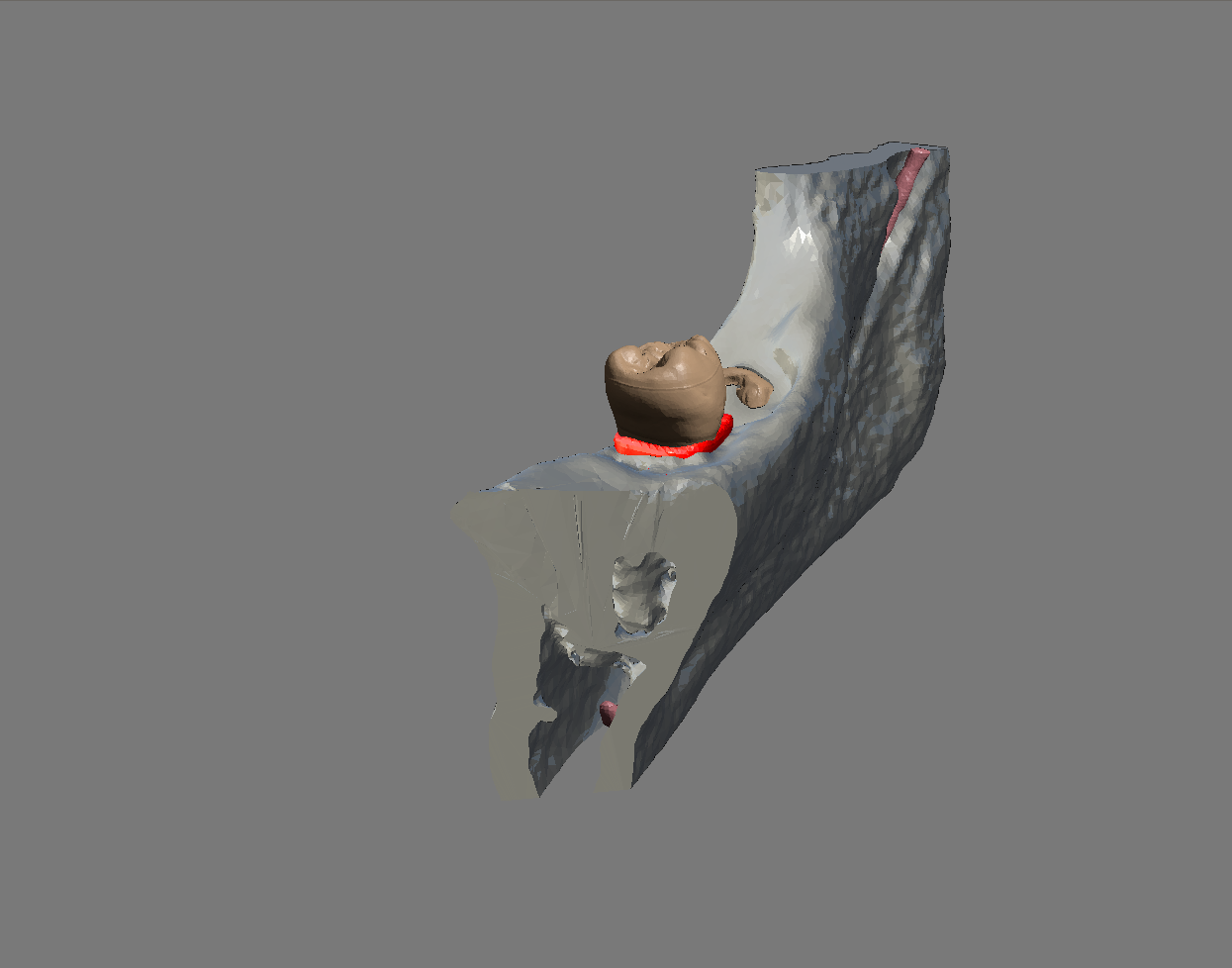 Image 18 of 19

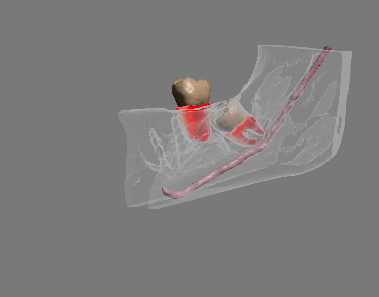 Image 19 of 19


Third Molar Extraction Model with Extractable Teeth and Alveolar Nerve Simulation
Overview
Elevate dental training with our innovative 3D Printed Third Molar Extraction Model. Designed to mimic real-life dental scenarios, this model is an essential tool for endodontists, periodontists, and implant specialists, offering a comprehensive and realistic training experience.
Key Features
Radiopaque Materials for Real-life Scanning and X-Ray Practice: Enhanced with radiopaque materials, our model provides an authentic experience for X-ray and scanning training, essential for mastering diagnostic imaging.
Proprietary Realistic Gum Tissue Material: Our unique gum tissue material offers a lifelike feel for suturing and flapping exercises, enriching the hands-on learning experience.
Medullary Bone Simulation for True-to-life Surgical Training: The inclusion of a simulated medullary bone adds a crucial element of realism, especially valuable for procedures involving bone work, like implant placement.
Two Extractable Teeth for Advanced Extraction Techniques: Featuring two extractable teeth, this model allows dental professionals to practice a variety of extraction methods, enhancing their skill set in dental surgery.
Alveolar Nerve with Simulated PDLs (Periodontal Ligaments): Our model includes a detailed simulation of the alveolar nerve with periodontal ligaments, offering an exceptional opportunity to understand and practice procedures involving nerve preservation and periodontal treatments.
Ideal for
Continuing Education (CE) Instructors
Dental and Endodontic Instrument Companies
Dental Schools and Training Institutions
Benefits
Comprehensive Skill Enhancement: From radiographic analysis to complex surgical procedures, our model covers an extensive range of dental practices.
Realistic and Hands-On Learning: Equipped with features like extractable teeth and simulated nerve structures, it offers an immersive training experience.
